|
The branchial (Gk.
gill) apparatus of a four-week-old
embryo consists of the branchial arches,
pouches, grooves (clefts), and membranes.
Each branchial arch (1, 2, 3, 4 and 6) is composed of lateral
mesoderm and neural crest cells
and each is associated with a cranial
nerve and an aortic arch.
Table 9
- Adult Derivatives of Pharyngeal Arches
|
|
|
Adult Derivatives
|
|
Arch
|
Nerve
|
Muscles (Mesoderm)
|
Skeletal Structures (Neural Crest)
|
|
First
(mandibular)
|
Trigeminal
(CN V)
|
Muscles of mastication, mylohyoid muscle
tensor veli palitini muscle, tensor tympani muscle, anterior belly of the
digastric muscle
|
Maxilla, zygomatic bone, temporal bone,
palatine bone, vomer, mandible, malleus, incus, sphenomandibular ligament
|
|
Second
(hyoid)
|
Facial
(CN VII)
|
Muscles of facial expression, stylohyoid
muscle, stapedius muscle posterior belly of digastric muscle
|
Stapes, styloid process, stylohyoid ligament,
lesser horn and superior body of the hyoid bone
|
|
Third
|
Glossopharyngeal
(CN IX)
|
Stylopharyngeus muscle
|
Greater horn and inferior body of the hyoid
bone
|
|
Fourth
|
Vagus
(CN X) – Superior laryngeal branch
|
Muscles of soft palate (except tensor veli
palatini) and muscles of pharynx (except stylopharyngeus), cricothyroid
muscle, cricopharyngeus muscle,
|
Thyroid cartilage, cricothyroid cartilage,
arytenoid cartilage, laryngeal cartilages
|
|
Sixth
|
Vagus
(CN X) –Recurrent laryngeal branch
|
Intrinsic muscles of the larynx (except
cricothyroid), upper (skeletal) muscles of esophagus
|
Laryngeal cartilages
|
Table 10
- Adult Derivatives of Pharyngeal Pouches
|
Pouch
|
Adult derivatives
|
|
1
|
Lining of auditory
tube and tympanic cavity
(middle ear cavity)
|
|
2
|
Largely obliterated, lining of intratonsillar cleft (tonsilar fossa)
|
|
3
|
Inferior
parathyroid glands, thymus
|
|
4
|
Superior
parathyroid glands, parafollicular
cells of thyroid gland
|

Figure
6 - Pharyngeal arches and pouches
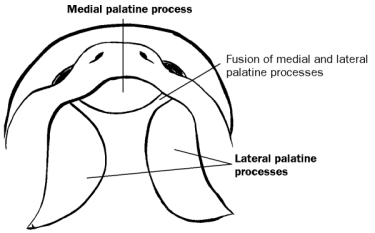
Figure 7 - Development of hard palate
Thyroid Gland
The thyroid gland begins as a downgrowth of the floor of
the pharynx called the thyroid
diverticulum. As it descends down the neck it remains connected to the
tongue via the thyroglossal duct. In
the adult a remnant of this duct persists in the tongue as the foramen cecum.
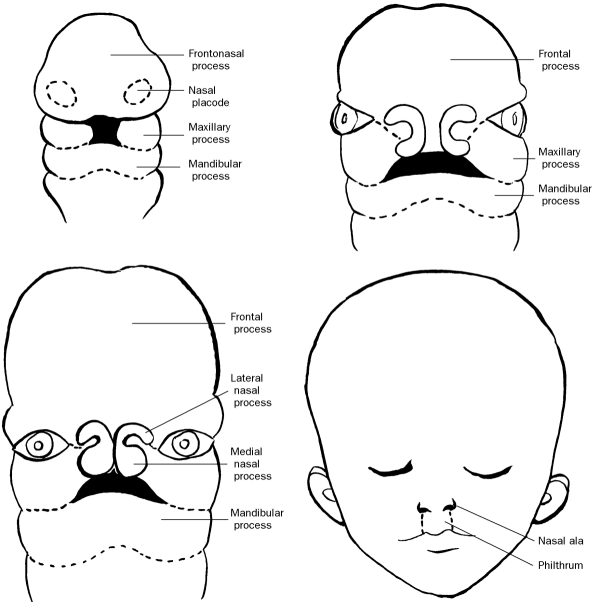
Figure 8 - Development of the face
|