The Normal Jugular Pulse (cont.)
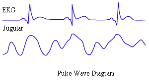
Instructions: Click on the portion of the diagram which is visible as an outward movement at the base of the neck. Begin with the most prominent waves.
-
CORRECT
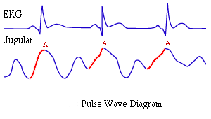
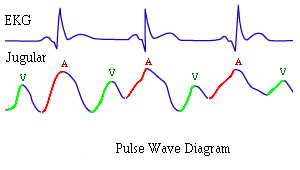
This is an A wave which is enhanced by right atrial contraction. Normally it's the largest and most easily visualized wave.
But there is more Try again.
-
CORRECT
This is an A wave which is enhanced by right atrial contraction. Normally it's the largest and most easily visualized wave.
But there is more Try again.
-
CORRECT
This is an A wave which is enhanced by right atrial contraction. Normally it's the largest and most easily visualized wave.
But there is more Try again.
-
INCORRECT
Select again.
-
CORRECT AGAIN
This is a V wave which is associated with the passive distension of the right atrium during ventricular systole. Normally it is small or not visible at all.
Note: Play the video one more time. Both the A and V waves are visible in this example.
Move on to the next screen.
-
CORRECT AGAIN
This is a V wave which is associated with the passive distension of the right atrium during ventricular systole. Normally it is small or not visible at all.
Note: Play the video one more time. Both the A and V waves are visible in this example.
Move on to the next screen.
-
CORRECT AGAIN
This is a V wave which is associated with the passive distension of the right atrium during ventricular systole. Normally it is small or not visible at all.
Note: Play the video one more time. Both the A and V waves are visible in this example.
Move on to the next screen.
-
CORRECT AGAIN
This is a V wave which is associated with the passive distension of the right atrium during ventricular systole. Normally it is small or not visible at all.
Note: Play the video one more time. Both the A and V waves are visible in this example.
Move on to the next screen.
-
INCORRECT
Note: Play the video one more time. Both the A and V waves are visible in this example.


