//
Summary:
Because of the patient's concern, an echo study was performed that showed no evidence of mitral valve abnormality.
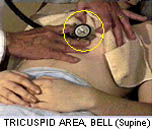
 Click
to review auscultation.
Click
to review auscultation.
Lessons:
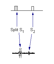
- Splitting of S1 is normal and must not be confused with the systolic click of mitral valve prolapse.
- Splitting of S1 is almost universal in the tricuspid area but is also often present at the apex in perfectly normal people.
- When in doubt have the patient squat.
a. A split S1 does not change.
b. A systolic click due to MVP moves later in systole and becomes more mid-systolic in cadence. - Review Case 4 on how to differentiate a split S1 from an S4.