Instructions: After reading the patient summary, click on the stethoscope head to listen to the heart sound. Then click on the diagram that best matches what you hear.
A 60-year-old police officer with recent onset of mild exertional dyspnea associated with moderate substernal discomfort. Quickly relieved by rest. Vitals: BP 116/80, P 76, R 12, Wt. 192, Ht. 72 inches PMH. Questionable rheumatic fever as a child. Known heart murmur for 20 years. FH. Negative for CV disease. SH. Smokes occasional cigar. Physical Examination: Slow-rising carotid upstrokes. Bilateral carotid bruits. No venous distension. Lungs clear. No cardiac enlargement. The apex impulse is sustained consistent with left ventricular hypertrophy. The following was present on auscultation.
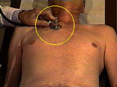
AORTIC AREA, DIAPHRAGM (Supine)
-
INCORRECT
This patient does not have a diastolic murmur. The heart sound matching this diagram is playing now. Please try again.
-
INCORRECT
This patient does not have an audible S2. The heart sound matching this diagram is playing now. Try again.
-
CORRECT
Play the phono and then, if you wish, click on the incorrect diagrams to compare heart sounds.
Move on to the next screen.