
|
|
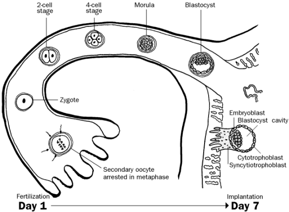
FertilizationAfter ovulation, the unfertilized egg is arrested in prophase of meiosis II and contains one polar body left over from meiosis I. Fertilization is a process of several events and typically takes place in the ampullated portion of the uterine tube: CapacitationChanges take place in the glycoprotein coat of sperm as they travel up the female reproductive tract. These changes are absolutely essential for fertilization. Thus, to perform successful in vitro fertilization you must add some tissue extracted from the female reproductive tract in addition to the sperm and egg extracted from the parents. ApproximationOnly a tiny fraction of sperm actually reaches the ampulla of the uterine tube to be near the egg. Penetration of Corona RadiataThe sperm uses both chemical and physical means to penetrate the egg’s corona radiata: · The action of membrane-bound enzyme hyaluronidase on its coat, and · Swimming motion of its flagellum. Penetration of Zona PellucidaOnce inside the corona radiata, the sperm binds to the species-specific ZP3 receptor on the egg’s glycoprotein coat. This triggers the acrosomal reaction, or the release of enzymes stored in the sperm’s acrosome (e.g. acrosin). These enzymes help the sperm “drill through” the zona pellucida. Once the sperm has penetrated the outer layers it fuses with the plasma membrane of the egg and releases its contents inside. The head and the tail of the sperm degrade, so that all mitochondria in the embryo (and all mitochondrial DNA) come from the mother. Cortical ReactionEntry of a sperm into the egg triggers changes that prevent polyspermy (fertilization of an egg by more than one sperm). These changes are known as the cortical reaction. Table 4 - Cortical Reaction
Fusion of PronucleiDNA in the male pronucleus is packed very tightly with protamines to make it compact enough to fit inside a sperm. These protamines are replaced by histones inside the egg, unpacking the DNA. Afterwards the male and female pronuclei fuse and the egg completes its second meiotic division, resulting in a second polar body. The fertilized egg is now known as the zygote (“together”). CleavageThe zygote undergoes a number of ordinary mitotic divisions that increase the number of cells in the zygote but not its overall size. Each cycle of division takes about 24 hours. The individual cells are known as blastomeres. At the 32-cell stage the embryo is known as a morula (L. “mulberry”), a solid ball consisting of an inner cell mass and an outer cell mass. The inner cell mass will eventually become the embryo and fetus, while the outer cell mass will eventually become part of the placenta. Blastocyst FormationCompactionThe cells on the outside of the morula form tight intercellular junctions and express ion channels to create an impermeable barrier. CavitationA fluid-filled cavity forms inside the morula. This cavity is known as the blastocyst cavity or blastocoele, and the morula is now called a blastula or blastocyst. The inner cell mass is now known as the embryoblast and the outer cell mass becomes the trophoblast. ImplantationHatchingThe blastula sheds its zona pellucida. This is required for implantation to occur. One function of the zona pellucida is to prevent premature implantation. Attachment and InvasionThe embryo attaches to and invades into the maternal
endometrium. The trophoblast
differentiates into the cytotrophoblast
and the syncytiotrophoblast.
The embryo typically implants in
the posterior superior wall of the uterus.
The response of the maternal endrometrial cells to the invading embryo is
called the decidual reaction. Figure 1 - Summary of the first week of development Clinical CorrelationsEctopic pregnancyThe bastocyst implants in a location other than the uterus. This can present as an acute surgical emergency for the mother after the fetus begins to outgrow its confines: Table 5 - Common Sites of Ectopic Pregnancy
Placenta PreviaThe embryo implants in the lower part of the uterus towards the cervix. This makes it easy for the placenta to tear, and the mother can die from hemorrhage, or the placenta may grow to obstruct the cervical canal. This is diagnosed with ultrasound, and the baby is delivered via Cesarean section.
|
|
|